
Endodontic treatment, known to patients as “root canal treatment”, is one of the most common dental procedures by which a tooth affected by deep decay, infection or trauma can be saved.
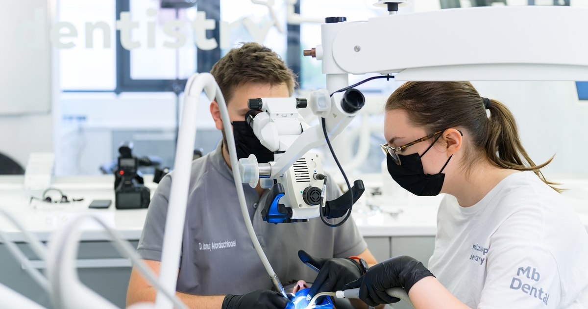
In recent years, the use of the dental operating microscope has significantly changed the way this type of treatment is carried out, increasing the precision of the intervention and the chances of long-term success. More and more specialized clinics have integrated this technology as a working standard in endodontic treatments.
What is endodontic treatment and when it becomes necessary
Root canal treatment involves removing the inflamed or infected tissue from inside the tooth, cleaning, disinfecting and sealing the root canals. Intervention becomes necessary when the infection reaches the level of the nerve, most often due to an untreated cavity or trauma.
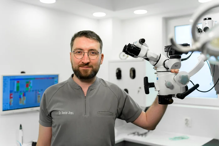
“Intense pain, persistent tenderness or inflammation are usually the signs that prompt patients to see a doctor“, he explains Dr. Stefan Jitarudentist in the MB Dental clinic, with a practice focused on endodontics and one of the first promoters of the use of the dental operating microscope in Cluj, for over 15 years.
One of the most widespread fears remains the association of root canal treatment with pain. In reality, modern procedures are performed under effective anesthesia, and discomfort is usually reduced.
The limits of classical treatment, without a microscope
In its classic form, root canal treatment is based on a limited view of the inside of the tooth. The root canal system is extremely fine and variable, and without magnification, certain details can easily be missed.
“Additional canals, calcifications or microcracks can go undetected without proper visualization, which directly influences treatment success,” explains Dr. Jitaru.
A frequent example is the MB2 canal, the second mesio-vestibular canal of the upper molars. Studies show that its identification rate is about 17% without magnification and exceeds 70% when the dental operating microscope is used.
Incomplete canal cleaning, failure to identify all anatomical structures, or poor sealing are among the most common causes of endodontic treatment failure.
What is the dental operating microscope and what does it change in practice
The dental operating microscope is optical equipment that provides controlled magnification and direct illumination of the operating field. In endodontics, it allows detailed visualization of the inside of the tooth so that each step of the procedure can be checked with precision.
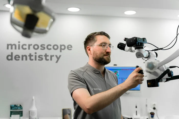
“The microscope fundamentally changes the way the doctor sees and treats the tooth. Basically, you work with a level of detail that is impossible to reach with the naked eye,” explains Dr. Ștefan Jitaru.
This approach allows for more conservative treatments with minimal removal of healthy tooth tissue and better control over each manipulation.
When the microscope becomes essential
The use of the dental operating microscope is particularly important in complex cases: endodontic retreatments, calcified canals, fractured instruments, perforations or atypical anatomies.
“In retreatments, the microscope helps us identify the exact cause of the initial failure and correct the problems that have arisen,” says Dr. Jitaru, a dentist with a practice focused on endodontics at the MB Dental clinic in Cluj.
Thanks to this technology, many teeth that would have been extracted in the past can today be saved and kept functional.
Direct benefits for the patient
For the patient, the main advantage of the treatment performed with a microscope is precision. A more thorough cleaning and sealing of the canals reduces the risk of reinfection, and subsequent restorations are more durable.
Systematic reviews show success rates of primary endodontic treatment ranging from 82% to 93%, and survival of treated teeth exceeding 90% at 10 years. A correctly adapted coronary restoration remains an essential factor in maintaining these long-term results.
In addition, patients usually report good comfort during the procedure and faster recovery due to more precise and less invasive interventions.
The dental operating microscope in Romania
Internationally, more than 90% of specialist endodontists use the dental operating microscope in their daily practice. In Romania, approximately 58% of dentists use some form of magnification, and nearly 28% use the dental operating microscope, with higher adoption in university centers and large cities.
The adoption of this technology was initially limited by the high costs of equipment, the need for dedicated training and changing the way of working. As the clinical benefits became apparent, more and more specialized centers integrated the microscope into daily practice.
The role of experience and continuing education
MB Dentalclinic in Cluj-Napoca and Târgu-Mureș, is one of the centers that early adopted the dental operating microscope and integrated it as a working standard in root canal treatments.
In addition to clinical activity, the team is also involved in the professional training of dentists, contributing to the promotion of modern endodontics and the correct use of the dental operating microscope.
What patients should know before a root canal
Specialists recommend that patients ask if the treatment is performed under a microscope, what experience the doctor has in endodontics and how the tooth will be restored after the treatment is completed.
“The final restoration is not a simple filling, but an essential stage for long-term success”, emphasizes Dr. Ștefan Jitaru.
Root canal treatment performed with a dental operating microscope, followed by a properly fitted restoration, can keep the tooth functional in the long term. Correct information and the choice of an experienced doctor remain key factors for a predictable and sustainable result.