
Despite decades of effort, scientists have never been able to recover dinosaur DNA. Most paleontological research today still focuses on looking for traces of original organic material in fossils, but DNA has not survived the passage of time.
Much of what we know about dinosaurs comes from fossilized bones and teeth. These durable remains are well preserved, but provide only limited information about how these animals actually lived, writes sciencedaily.com.
Soft tissues, on the other hand, can reveal much more. These rare fossilized materials include muscles and ligaments, pigments, or even skin (such as scales or feathers). They provide important clues about appearance, movement and behavior.
Another type of soft tissue, sometimes preserved inside bones, is blood vessels. The research team identified preserved blood vessels in a Tyrannosaurus rex fossil, and the findings were recently published in Scientific Reports.
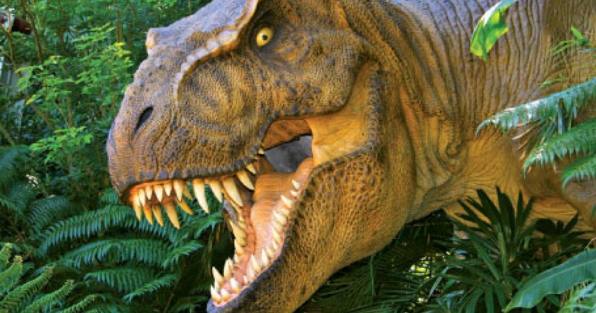
The largest T-Rex ever found
The preserved vessels analyzed by the research team come from an extraordinary specimen known as Scotty. Housed at the Royal Saskatchewan Museum in Canada, Scotty is the largest T. rex ever discovered and one of the most complete.
Evidence suggests that Scotty lived a difficult life about 66 million years ago. Many of its bones show signs of injury, possibly from a fight with another dinosaur or from disease. A rib sticks out, showing a large fracture that had only partially healed.
When bones are damaged, the body increases the activity of blood vessels in the affected area to support healing. The structures the researchers observed in Scotty’s coast appear to be part of this process, forming a dense network of mineralized vessels, which we reconstructed using 3D models.
Advanced imaging reveals hidden structures
Studying the interior of fossil bones presents two major challenges. First, researchers must look inside without damaging the specimen. Second, fossilized bones are extremely dense because minerals have replaced the original organic material over millions of years.
Initially, the researchers considered using a computed tomography (CT) scan, similar to those used in medicine. Although this method is non-destructive, standard CT scanners cannot penetrate the dense structure of large fossils.
The researchers then turned to synchrotron light, a powerful form of high-intensity X-rays produced in specialized particle accelerator facilities. This technique allowed them to visualize small internal features such as blood vessels with remarkable clarity.
Synchrotron imaging also allowed analysis of the chemical composition of the structures. The vessels were preserved as iron-rich mineralized casts, which is a common fossilization process.
What Blood Vessels Reveal About Dinosaur Life
The partially healed fracture in Scotty’s rib provides a rare opportunity to study how a T. rex recovered from a wound. By examining preserved blood vessels, researchers can gain insights into healing processes and survival strategies in large predatory dinosaurs.
The findings could also guide future fossil research. Bones that show signs of injury or disease may be more likely to preserve blood vessels or other soft tissue, helping scientists identify promising specimens.
By combining physics, paleontology, and advanced imaging technologies, researchers are beginning to uncover details about dinosaur biology that were once thought impossible to study.